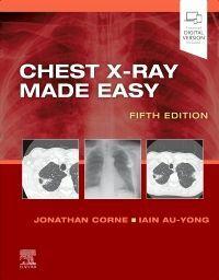























책 이미지

eBook 미리보기
책 정보
· 제목 : 흉부 X선 영상 간편 판독
· 분류 : 국내도서 > 대학교재/전문서적 > 의약학간호계열 > 임상의학 > 영상의학과
· ISBN : 9791124176016
· 쪽수 : 576쪽
· 출판일 : 2026-01-26
· 분류 : 국내도서 > 대학교재/전문서적 > 의약학간호계열 > 임상의학 > 영상의학과
· ISBN : 9791124176016
· 쪽수 : 576쪽
· 출판일 : 2026-01-26
책 소개
초급 의사와 의대생을 위한 흉부 X선 교재로, 흉부 X선 촬영을 최대한 쉽게 판독할 수 있도록 고안되었다. 단순히 방사선학적 소견을 설명하는 것이 아니라, 임상 현장에서 흔히 접할 수 있는 이상 소견을 이해하는...
흉부 X선 촬영은 병원에서 가장 흔히 시행되는 검사이다. 초급 의사와 의대생을 위한 교재로, 흉부 X선 촬영을 최대한 쉽게 판독할 수 있도록 고안되었다. 단순히 방사선학적 소견을 설명하는 것이 아니라, 임상 현장에서 흔히 접할 수 있는 이상 소견을 이해하는데 도움이 되는 안내서이다. 제5판에서는 내용을 확장하고 더욱 고품질의 최신 영상을 추가하였다.
목차
1. 흉부영상 촬영 시기와 방법 무엇을, 언제, 어떻게 처방할까요?
흉부 X선 검사 처방
흉부 X선 처방 후 처리 과정
2. 흉부 X선 촬영 영상 판독법
기본 판독은 쉽다
촬영 영상의 기술적 품질
후전방(PA) 영상
측면 영상 판독방법
3. 전산화단층촬영(The CT scan)
전산화단층촬영(CT scanning)
전산화단층촬영 유형(Types of CT scan)
영상 판독(Interpreting the images)
흉부 전산화단층촬영 판독법
인공 음영(Artefacts)
4. 측면 흉부 영상(The lateral chest x-ray)
폐 영상(The lungs)
심장 영상(The heart)
5. 백색 폐 음영(The white lung field)
폐 허탈(Collapse)
폐용적 감소(Volume loss)
폐 경결(Consolidation)
주폐포자충 폐렴(Pneumocystis carinii (jiroveci) pneumonia, PCP)
흉막 삼출(Pleural effusion)
흉막판(Pleural plaques)[석면판 (Asbestos plaques]
중피종(Mesothelioma)
흉부 전산화단층촬영에서 흉막 질환(Pleural disease on a CT scan)
폐결절(Lung nodule)
공동성 폐 질환(Cavitating lung lesion)
좌심부전(Left ventricular failure, LVF)
급성호흡곤란증후군(ARDS)
기관지 확장증(Bronchiectasis)
섬유증(Fibrosis)
수두 폐렴(Chickenpox pneumonia)
속립성 음영(Miliary shadowing)
6. 흑색 폐야(The black lung field)
만성폐쇄성폐질환(COPD)
기흉(Pneumothorax)
긴장성 기흉(Tension pneumothorax)
폐색전증(Pulmonary Embolus, PE)
유방절제술(Mastectomy)
7. 이상 폐문(The abnormal hilum)
일측성 폐문 비대(Unilateral hilar enlargement)
양측성 폐문 비대(Bilateral hilar enlargement)
8. 심장 이상 음영(The abnormal heart shadow)
심낭 삼출(Pericardial effusion)
심방중격결손(Atrial septal defect, ASD)
승모판 협착(Mitral stenosis)
좌심실 동맥류(Left ventricular aneurysm)
9. 종격동 확장(The widened mediastinum)
10. 늑골 이상(Abnormal ribs)
늑골 골절(Rib fractures)
전이성 침착(Metastatic deposits)
11. 연조직 이상(Abnormal soft tissues)
수술적 폐기종(Surgical emphysema)
12. 중증 환자(The sick patient)
라인, 튜브, 장치(Lines, tubes and devices)
중환자실 환자(The patient on intensive care)
13. 감춰진 이상(The hidden abnormality)
판코스트 종양(Pancoast tumour)
심장 뒤쪽 이상 음영(Abnormalities behind the heart)
횡격막 아래 공기 음염(Air under the diaphragm)
14. 흉부 영상 퀴즈(The chest x-ray quiz)
흉부 X선 검사 처방
흉부 X선 처방 후 처리 과정
2. 흉부 X선 촬영 영상 판독법
기본 판독은 쉽다
촬영 영상의 기술적 품질
후전방(PA) 영상
측면 영상 판독방법
3. 전산화단층촬영(The CT scan)
전산화단층촬영(CT scanning)
전산화단층촬영 유형(Types of CT scan)
영상 판독(Interpreting the images)
흉부 전산화단층촬영 판독법
인공 음영(Artefacts)
4. 측면 흉부 영상(The lateral chest x-ray)
폐 영상(The lungs)
심장 영상(The heart)
5. 백색 폐 음영(The white lung field)
폐 허탈(Collapse)
폐용적 감소(Volume loss)
폐 경결(Consolidation)
주폐포자충 폐렴(Pneumocystis carinii (jiroveci) pneumonia, PCP)
흉막 삼출(Pleural effusion)
흉막판(Pleural plaques)[석면판 (Asbestos plaques]
중피종(Mesothelioma)
흉부 전산화단층촬영에서 흉막 질환(Pleural disease on a CT scan)
폐결절(Lung nodule)
공동성 폐 질환(Cavitating lung lesion)
좌심부전(Left ventricular failure, LVF)
급성호흡곤란증후군(ARDS)
기관지 확장증(Bronchiectasis)
섬유증(Fibrosis)
수두 폐렴(Chickenpox pneumonia)
속립성 음영(Miliary shadowing)
6. 흑색 폐야(The black lung field)
만성폐쇄성폐질환(COPD)
기흉(Pneumothorax)
긴장성 기흉(Tension pneumothorax)
폐색전증(Pulmonary Embolus, PE)
유방절제술(Mastectomy)
7. 이상 폐문(The abnormal hilum)
일측성 폐문 비대(Unilateral hilar enlargement)
양측성 폐문 비대(Bilateral hilar enlargement)
8. 심장 이상 음영(The abnormal heart shadow)
심낭 삼출(Pericardial effusion)
심방중격결손(Atrial septal defect, ASD)
승모판 협착(Mitral stenosis)
좌심실 동맥류(Left ventricular aneurysm)
9. 종격동 확장(The widened mediastinum)
10. 늑골 이상(Abnormal ribs)
늑골 골절(Rib fractures)
전이성 침착(Metastatic deposits)
11. 연조직 이상(Abnormal soft tissues)
수술적 폐기종(Surgical emphysema)
12. 중증 환자(The sick patient)
라인, 튜브, 장치(Lines, tubes and devices)
중환자실 환자(The patient on intensive care)
13. 감춰진 이상(The hidden abnormality)
판코스트 종양(Pancoast tumour)
심장 뒤쪽 이상 음영(Abnormalities behind the heart)
횡격막 아래 공기 음염(Air under the diaphragm)
14. 흉부 영상 퀴즈(The chest x-ray quiz)